TriTom Imaging Platform
Small Animal Whole Body In Vivo Imaging based on PhotoAcoustic Fluorescence Tomography (PAFT)
- Overview
- Features
- Technical Details
- Application
- Download
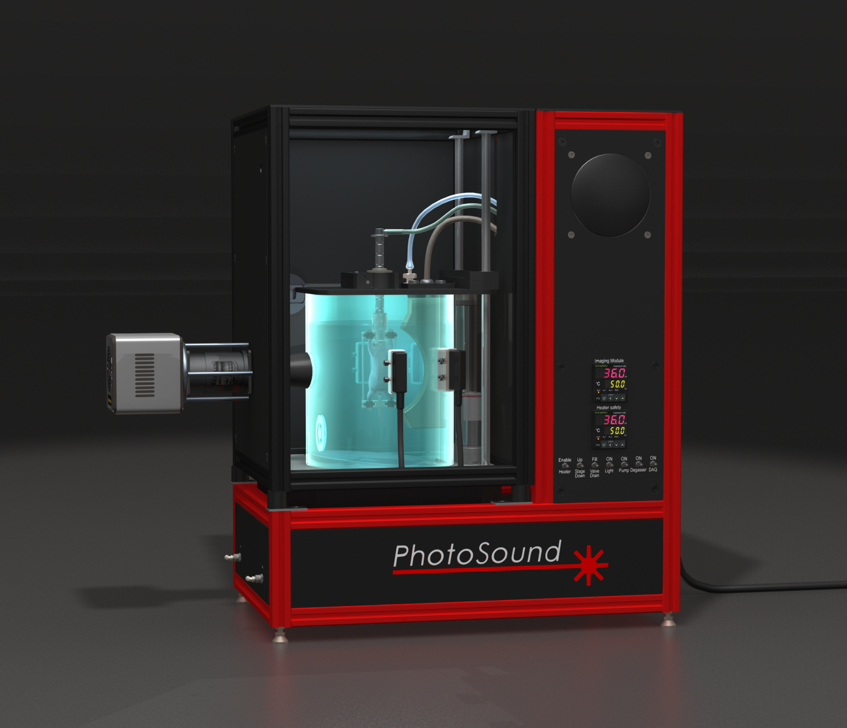
The patented TriTom imaging platform is based on Photoacoustic Fluorescence Tomography (PAFT) technology that provides unparalleled capabilities for whole body imaging and in vivo characterization of small animal models. Complimentary 3D imaging modalities are integrated into a single powerful instrument by enabling co-registered Photoacoustic Tomography (PAT) and Fluorescence Molecular Tomography (FMT). Utilizing an innovative, compact configuration, simultaneous co-registered collection of orthogonal photoacoustic and optical projections can be acquired. The platform provides high-resolution robust anatomical registration of optical biomarkers, while maintaining high molecular sensitivity. A broad spectrum of preclinical research applications include cancer, toxicology, tissue engineering and regeneration, cardiovascular and developmental biology.
TriTom Imaging System Introductory Video
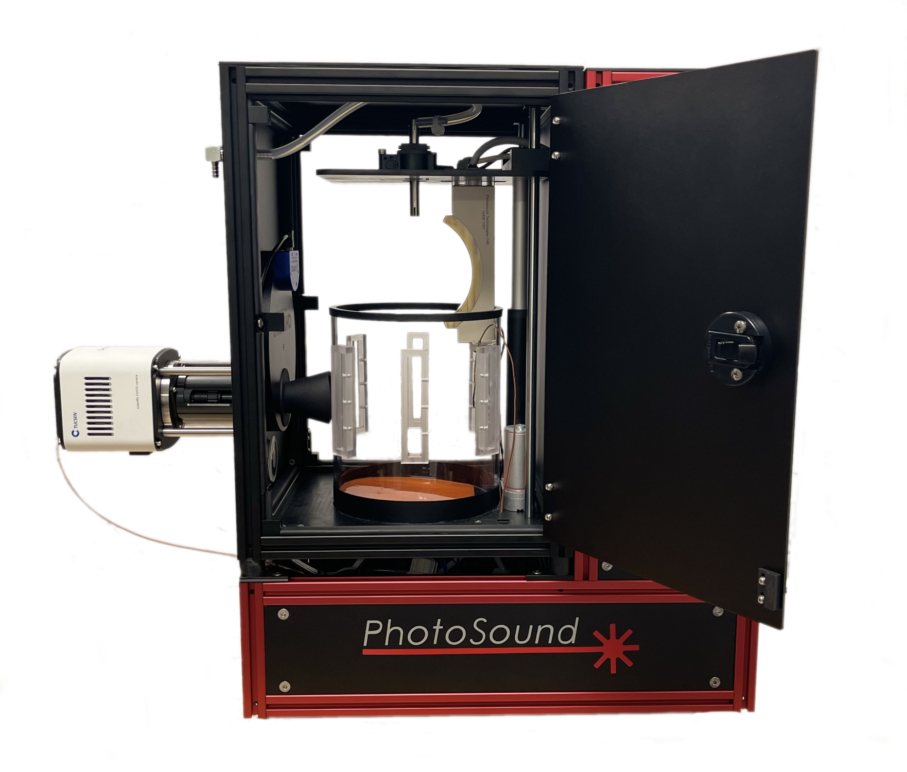
- Imaging chamber with optimally arranged optical excitation ports and safety interlocks on dual access doors
- 96-channel data acquisition unit optimized for detection of photoacoustic waves
- EMI protected 96-channel curved transducer array
- sCMOS camera covering a broad range of fluorescent and bioluminescent emission
- Temperature control unit maintains sample environment within ± 0.1°C
- Water control unit maintains clean, heated sound coupling medium with fill/drain/degassing
- Durable key slot extrusion housing with safety interlocks
- Precision rotary stage enabling tomographic scan of the sample
- Built-in life support delivers oxygen/anesthesia directly to the sample
Technical Specifications
- 36 sec single excitation wavelength scan duration
- 3D PAT reconstruction time: <30 secs
- 150 µm (x & y axes) 500 µm (z axis) PAT resolution
- 16 line pairs/mm FLT resolution
- µa10 = 0.1 cm-1 PAT sensitivity (equals ~1 µM ICG in water)
- > 30,000 virtual channels
- 30 x 30 x 30 mm visualization volume
-
Surface Photoacoustic Tomography
-
Volumetric Deep Body Photoacoustic Tomography
-
Fluorescence Tomography