- Overview
- Features
- Technical Details
- Application
- Download
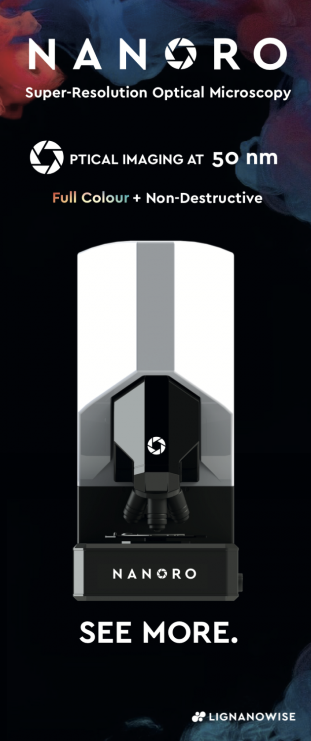
A NEW WAY OF SEEING
Super-resolution Microsphere Amplifying Lens: SMAL
NANORO M has redefined the smallest object that the eye can see, using the most popular form of imaging in the world: white light, optical microscopy. Our super-resolution technique pushes light past its theoretical limit, using microsphere optics to break the diffraction limit of light. We want to democratise super-resolution, making it an accessible, easy to imaging technique, that doesn't require a clean room, a huge power supply, complex sample preparation, or significant training. The super-resolution microscopes are not prohibitively expensive. Whether you are a university, a start-up, or a multi-national it is our mission to make super-resolution imaging accessible to everyone. Because the microscopes are purely optical they are non-destructive, they work with both metallic on and non-metallic samples, they are easy to use and resolve down to 80 nm (on certain sample types) in full, real, colour.

Full Colour
Our microsphere lenses collect sub-diffraction information and project it into the far-field. Not only do they see in super-resolution, but in full colour.

NON-DESTRUCTIVE
Unlike conventional super-resolution techniques our microscopes are none-destructive. Allowing you to image your sample multiple times without compromising its integrity.

ACCESSIBLE TO ALL
Because our microscopes are fully optical with super-resolution capability, they are easy to use for anyone familiar with conventional white-light microscopy.

POWERFUL SOFTWARE
Our software suite allows you to rapidly image large areas of a sample at multiple resolutions, and with our one click export you can then easily feed your images to Image

DOWN TO 80 NM
With the option of using SMAL, our microscopes can resolve lateral features down to 80 nm depending on the sample.

LAB READY
Our microscopes work without the need for prohibitive environmental conditions. They work on a standard optical table requiring no clean room, vacuum, etc.
Technical Specifications

Graphene Imaging
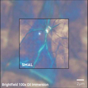
On the left is an example of SMAL imaging vs. brightfield optical microscopy with a 100x oil immersion lens. Note the dramatic increase in both resolution and magnification.

50 nm Lateral resolution
Here you can see how the NANOPSIS M is able to resolve lateral gaps of 50 nm, far beyond the reaches of standard optical microscopy.

Semiconductor Imaging
Here you can see a comparison between standard optical microscopy and SMAL microscopy while observing Semiconductor samples